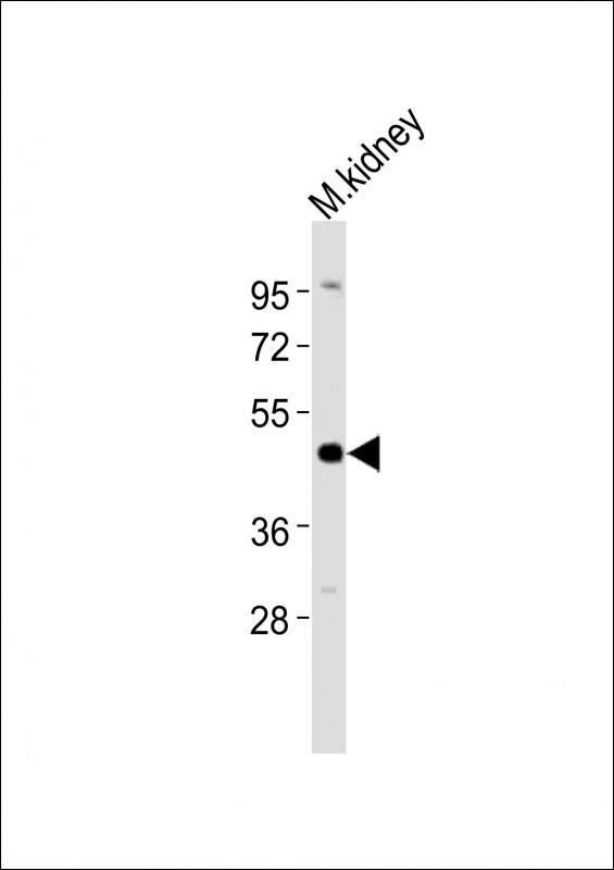
Anti-Ihh Antibody (N-term) at 1:2000 dilution + mouse kidney lysates
Lysates/proteins at 20 μg per lane.
Secondary
Goat Anti-Rabbit IgG, (H+L), Peroxidase conjugated at 1/10000 dilution
Predicted band size : 45 kDa
Blocking/Dilution buffer: 5% NFDM/TBST.

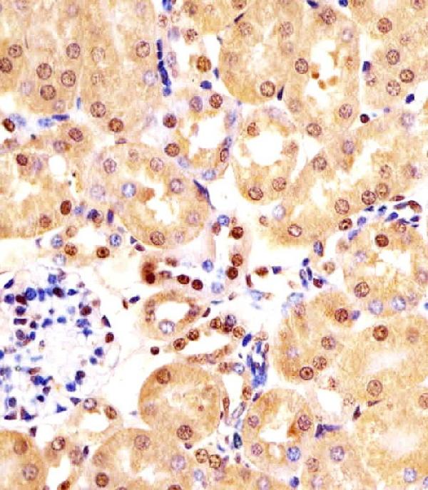
M01448 staining (Mouse) Ihh in mouse kidney sections by Immunohistochemistry (IHC-P -paraformaldehyde-fixed, paraffin-embedded sections). Tissue was fixed with formaldehyde and blocked with 3% BSA for 0. 5 hour at room temperature; antigen retrieval was by heat mediation with a citrate buffer (pH6). Samples were incubated with primary antibody (1/25) for 1 hours at 37~C. A undiluted biotinylated goat polyvalent antibody was used as the secondary antibody.

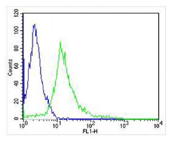
Overlay histogram showing Jurkat cells stained with M01448 (green line). The cells were fixed with 4% paraformaldehyde (10 min) and then permeabilized with 90% methanol for 10 min. The cells were then icubated in 2% bovine serum albumin to block non-specific protein-protein interactions followed by the antibody (KRT12 Antibody, 1:25 dilution) for 60 min at 37oC. The secondary antibody used was Alexa Fluor? 488 goat anti-rabbit lgG (H+L) at 1/400 dilution for 40 min at 37oC. Isotype control antibody (blue line) was rabbit IgG1 (1g/1x10^6 cells) used under the same conditions. Acquisition of >10, 000 events was performed.

Anti-Ihh Antibody (N-term) at 1:2000 dilution + mouse kidney lysates
Lysates/proteins at 20 μg per lane.
Secondary
Goat Anti-Rabbit IgG, (H+L), Peroxidase conjugated at 1/10000 dilution
Predicted band size : 45 kDa
Blocking/Dilution buffer: 5% NFDM/TBST.

M01448 staining (Mouse) Ihh in mouse kidney sections by Immunohistochemistry (IHC-P -paraformaldehyde-fixed, paraffin-embedded sections). Tissue was fixed with formaldehyde and blocked with 3% BSA for 0. 5 hour at room temperature; antigen retrieval was by heat mediation with a citrate buffer (pH6). Samples were incubated with primary antibody (1/25) for 1 hours at 37~C. A undiluted biotinylated goat polyvalent antibody was used as the secondary antibody.

Overlay histogram showing Jurkat cells stained with M01448 (green line). The cells were fixed with 4% paraformaldehyde (10 min) and then permeabilized with 90% methanol for 10 min. The cells were then icubated in 2% bovine serum albumin to block non-specific protein-protein interactions followed by the antibody (KRT12 Antibody, 1:25 dilution) for 60 min at 37oC. The secondary antibody used was Alexa Fluor? 488 goat anti-rabbit lgG (H+L) at 1/400 dilution for 40 min at 37oC. Isotype control antibody (blue line) was rabbit IgG1 (1g/1x10^6 cells) used under the same conditions. Acquisition of >10, 000 events was performed.


