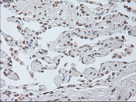
Figure 1. Immunohistochemical staining of paraffin-embedded Human lung tissue within the normal limits using anti-IL-6 mouse monoclonal antibody. (Heat-induced epitope retrieval by 10mM citric buffer, pH6.0, 100°C for 10min, MA00102)
all(19)
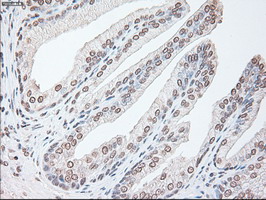
Figure 2. Immunohistochemical staining of paraffin-embedded Human prostate tissue within the normal limits using anti-IL-6 mouse monoclonal antibody. (Heat-induced epitope retrieval by 10mM citric buffer, pH6.0, 100°C for 10min, MA00102)
all(19) 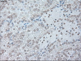
Figure 3. Immunohistochemical staining of paraffin-embedded Carcinoma of Human kidney tissue using anti-IL-6 mouse monoclonal antibody. (Heat-induced epitope retrieval by 10mM citric buffer, pH6.0, 100°C for 10min, MA00102)
all(19) 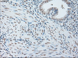
Figure 4. Immunohistochemical staining of paraffin-embedded Human endometrium tissue within the normal limits using anti-IL-6 mouse monoclonal antibody. (Heat-induced epitope retrieval by 10mM citric buffer, pH6.0, 100°C for 10min, MA00102)
all(19) 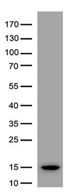
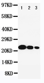
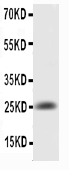
Figure 5. Western blot analysis of recombinant IL6 protein (0.02ug) by using anti-IL6 antibody (Cat# MA00102).
all(19) 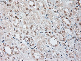
Figure 6. Immunohistochemical staining of paraffin-embedded Human Kidney tissue within the normal limits using anti-IL-6 mouse monoclonal antibody. (Heat-induced epitope retrieval by 10mM citric buffer, pH6.0, 100°C for 10min, MA00102)
all(19) 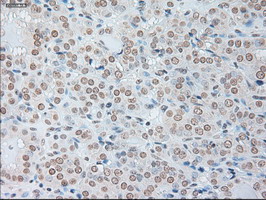
Figure 7. Immunohistochemical staining of paraffin-embedded Carcinoma of Human thyroid tissue using anti-IL-6 mouse monoclonal antibody. (Heat-induced epitope retrieval by 10mM citric buffer, pH6.0, 100°C for 10min, MA00102)
all(19) 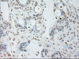
Figure 8. Immunohistochemical staining of paraffin-embedded Adenocarcinoma of Human colon tissue using anti-IL-6 mouse monoclonal antibody. (Heat-induced epitope retrieval by 10mM citric buffer, pH6.0, 100°C for 10min, MA00102)
all(19) 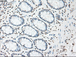
Figure 9. Immunohistochemical staining of paraffin-embedded Human colon tissue within the normal limits using anti-IL-6 mouse monoclonal antibody. (Heat-induced epitope retrieval by 10mM citric buffer, pH6.0, 100°C for 10min, MA00102)
all(19) 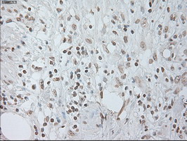
Figure 10. Immunohistochemical staining of paraffin-embedded Carcinoma of Human pancreas tissue using anti-IL-6 mouse monoclonal antibody. (Heat-induced epitope retrieval by 10mM citric buffer, pH6.0, 100°C for 10min, MA00102)
all(19) 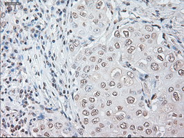
Figure 11. Immunohistochemical staining of paraffin-embedded Adenocarcinoma of Human breast tissue using anti-IL-6 mouse monoclonal antibody. (Heat-induced epitope retrieval by 10mM citric buffer, pH6.0, 100°C for 10min, MA00102)
all(19) 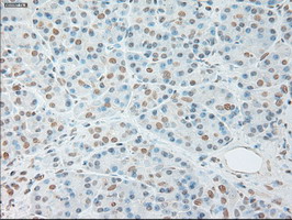
Figure 12. Immunohistochemical staining of paraffin-embedded Human pancreas tissue within the normal limits using anti-IL-6 mouse monoclonal antibody. (Heat-induced epitope retrieval by 10mM citric buffer, pH6.0, 100°C for 10min, MA00102)
all(19) 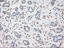
Figure 13. Immunohistochemical staining of paraffin-embedded Human breast tissue within the normal limits using anti-IL-6 mouse monoclonal antibody. (Heat-induced epitope retrieval by 10mM citric buffer, pH6.0, 100°C for 10min, MA00102)
all(19) 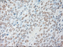
Figure 14. Immunohistochemical staining of paraffin-embedded Adenocarcinoma of Human ovary tissue using anti-IL-6 mouse monoclonal antibody. (Heat-induced epitope retrieval by 10mM citric buffer, pH6.0, 100°C for 10min, MA00102)
all(19) 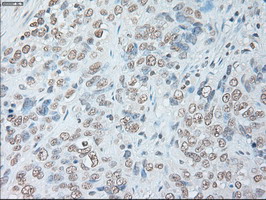
Figure 15. Immunohistochemical staining of paraffin-embedded Carcinoma of Human bladder tissue using anti-IL-6 mouse monoclonal antibody. (Heat-induced epitope retrieval by 10mM citric buffer, pH6.0, 100°C for 10min, MA00102)
all(19) 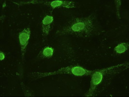
Figure 16. Anti-IL-6 mouse monoclonal antibody immunofluorescent staining of COS7 cells transiently transfected by pCMV6-ENTRY IL-6 .
all(19) 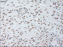
Figure 17. Immunohistochemical staining of paraffin-embedded Human Ovary tissue within the normal limits using anti-IL-6 mouse monoclonal antibody. (Heat-induced epitope retrieval by 10mM citric buffer, pH6.0, 100°C for 10min, MA00102)
all(19) 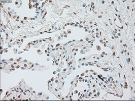
Figure 18. Immunohistochemical staining of paraffin-embedded Carcinoma of Human lung tissue using anti-IL-6 mouse monoclonal antibody. (Heat-induced epitope retrieval by 10mM citric buffer, pH6.0, 100°C for 10min, MA00102)
all(19) 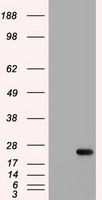
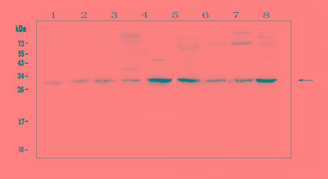
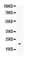
Figure 19. HEK293T cells were transfected with the pCMV6-ENTRY control (Left lane) or pCMV6-ENTRY IL-6 (Right lane) cDNA for 48 hrs and lysed. Equivalent amounts of cell lysates (5 ug per lane) were separated by SDS-PAGE and immunoblotted with anti-IL-6 (Cat# MA00102).
all(19) | Western blot (WB): | 1:1000 |
| Immunohistochemistry (IHC): | 1:50 |
| Immunofluorescence (IF): | 1:100 |
Figure 1. Immunohistochemical staining of paraffin-embedded Human lung tissue within the normal limits using anti-IL-6 mouse monoclonal antibody. (Heat-induced epitope retrieval by 10mM citric buffer, pH6.0, 100°C for 10min, MA00102)
Figure 2. Immunohistochemical staining of paraffin-embedded Human prostate tissue within the normal limits using anti-IL-6 mouse monoclonal antibody. (Heat-induced epitope retrieval by 10mM citric buffer, pH6.0, 100°C for 10min, MA00102)
Figure 3. Immunohistochemical staining of paraffin-embedded Carcinoma of Human kidney tissue using anti-IL-6 mouse monoclonal antibody. (Heat-induced epitope retrieval by 10mM citric buffer, pH6.0, 100°C for 10min, MA00102)
Figure 4. Immunohistochemical staining of paraffin-embedded Human endometrium tissue within the normal limits using anti-IL-6 mouse monoclonal antibody. (Heat-induced epitope retrieval by 10mM citric buffer, pH6.0, 100°C for 10min, MA00102)
Figure 5. Western blot analysis of recombinant IL6 protein (0.02ug) by using anti-IL6 antibody (Cat# MA00102).
Figure 6. Immunohistochemical staining of paraffin-embedded Human Kidney tissue within the normal limits using anti-IL-6 mouse monoclonal antibody. (Heat-induced epitope retrieval by 10mM citric buffer, pH6.0, 100°C for 10min, MA00102)
Figure 7. Immunohistochemical staining of paraffin-embedded Carcinoma of Human thyroid tissue using anti-IL-6 mouse monoclonal antibody. (Heat-induced epitope retrieval by 10mM citric buffer, pH6.0, 100°C for 10min, MA00102)
Figure 8. Immunohistochemical staining of paraffin-embedded Adenocarcinoma of Human colon tissue using anti-IL-6 mouse monoclonal antibody. (Heat-induced epitope retrieval by 10mM citric buffer, pH6.0, 100°C for 10min, MA00102)
Figure 9. Immunohistochemical staining of paraffin-embedded Human colon tissue within the normal limits using anti-IL-6 mouse monoclonal antibody. (Heat-induced epitope retrieval by 10mM citric buffer, pH6.0, 100°C for 10min, MA00102)
Figure 10. Immunohistochemical staining of paraffin-embedded Carcinoma of Human pancreas tissue using anti-IL-6 mouse monoclonal antibody. (Heat-induced epitope retrieval by 10mM citric buffer, pH6.0, 100°C for 10min, MA00102)
Figure 11. Immunohistochemical staining of paraffin-embedded Adenocarcinoma of Human breast tissue using anti-IL-6 mouse monoclonal antibody. (Heat-induced epitope retrieval by 10mM citric buffer, pH6.0, 100°C for 10min, MA00102)
Figure 12. Immunohistochemical staining of paraffin-embedded Human pancreas tissue within the normal limits using anti-IL-6 mouse monoclonal antibody. (Heat-induced epitope retrieval by 10mM citric buffer, pH6.0, 100°C for 10min, MA00102)
Figure 13. Immunohistochemical staining of paraffin-embedded Human breast tissue within the normal limits using anti-IL-6 mouse monoclonal antibody. (Heat-induced epitope retrieval by 10mM citric buffer, pH6.0, 100°C for 10min, MA00102)
Figure 14. Immunohistochemical staining of paraffin-embedded Adenocarcinoma of Human ovary tissue using anti-IL-6 mouse monoclonal antibody. (Heat-induced epitope retrieval by 10mM citric buffer, pH6.0, 100°C for 10min, MA00102)
Figure 15. Immunohistochemical staining of paraffin-embedded Carcinoma of Human bladder tissue using anti-IL-6 mouse monoclonal antibody. (Heat-induced epitope retrieval by 10mM citric buffer, pH6.0, 100°C for 10min, MA00102)
Figure 16. Anti-IL-6 mouse monoclonal antibody immunofluorescent staining of COS7 cells transiently transfected by pCMV6-ENTRY IL-6 .
Figure 17. Immunohistochemical staining of paraffin-embedded Human Ovary tissue within the normal limits using anti-IL-6 mouse monoclonal antibody. (Heat-induced epitope retrieval by 10mM citric buffer, pH6.0, 100°C for 10min, MA00102)
Figure 18. Immunohistochemical staining of paraffin-embedded Carcinoma of Human lung tissue using anti-IL-6 mouse monoclonal antibody. (Heat-induced epitope retrieval by 10mM citric buffer, pH6.0, 100°C for 10min, MA00102)
Figure 19. HEK293T cells were transfected with the pCMV6-ENTRY control (Left lane) or pCMV6-ENTRY IL-6 (Right lane) cDNA for 48 hrs and lysed. Equivalent amounts of cell lysates (5 ug per lane) were separated by SDS-PAGE and immunoblotted with anti-IL-6 (Cat# MA00102).
 联系我们
联系我们027-67845390
 关注我们
关注我们
本司产品仅用于科研,不用于临床诊断和治疗
联系方式:027-67845390/1/2 技术支持:武汉丰网
© 1993-2025 Boster Biological Technology co.Itd E-mail:boster@boster.com
鄂ICP备05005548号-2
 鄂公网安备 42018502007312号

 积分商城
积分商城  购物车
购物车  登录/注册
登录/注册  您当前的位置:
  说明书
说明书 一键复制产品信息

 成功添加到购物车
 微信客服
微信客服


 微信扫一扫立即咨询
