| Western blot (WB): | 1:3000-10000 |
| IHC : | 1:100-1:200 |
| Immunocytochemistry/Immunofluorescence (ICC/IF): | 1:100-200 |

Western blot analysis of anti-Beta Actin/ACTB antibody (M01263-4). The sample well of each lane was loaded with 30 ug of sample under reducing conditions.
Lane 1: human Hela whole cell lysates,
Lane 2: human K562 whole cell lysates,
Lane 3: pig brain tissue lysates.
After electrophoresis, proteins were transferred to a membrane. Then the membrane was incubated with mouse anti-Beta Actin/ACTB antigen affinity purified monoclonal antibody (M01263-4) at a dilution of 1:1000 and probed with a goat anti-mouse IgG-HRP secondary antibody (Catalog # BA1050). The signal is developed using ECL Plus Western Blotting Substrate (Catalog # AR1197). A specific band was detected for Beta Actin/ACTB at approximately 42 kDa. The expected band size for Beta Actin/ACTB is at 42 kDa.

Western blot analysis of anti-Beta Actin/ACTB antibody (M01263-4). The sample well of each lane was loaded with 30 ug of sample under reducing conditions.
Lane 1: rat brain tissue lysates,
Lane 2: rat liver tissue lysates,
Lane 3: rat NRK whole cell lysates,
Lane 4: mouse brain tissue lysates,
Lane 5: mouse NIH/3T3 whole cell lysates,
Lane 6: chicken liver tissue lysates,
Lane 7: chicken brain tissue lysates,
After electrophoresis, proteins were transferred to a membrane. Then the membrane was incubated with mouse anti-Beta Actin/ACTB antigen affinity purified monoclonal antibody (M01263-4) at a dilution of 1:1000 and probed with a goat anti-mouse IgG-HRP secondary antibody (Catalog # BA1050). The signal is developed using ECL Plus Western Blotting Substrate (Catalog # AR1197). A specific band was detected for Beta Actin/ACTB at approximately 42 kDa. The expected band size for Beta Actin/ACTB is at 42 kDa.

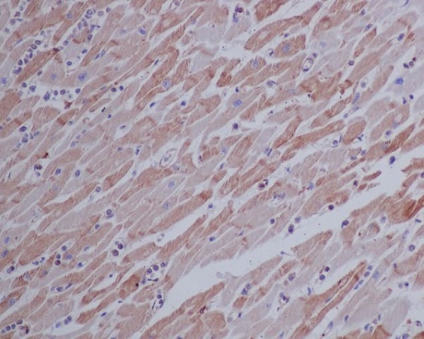
Immunohistochemical analysis of paraffin-embedded human heart, using beta Actin Mouse Monoclonal Antibody.

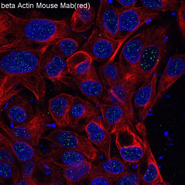
Immunofluorescent analysis of HepG2 cells, using beta Actin mAb.

Western blot analysis of anti-Beta Actin/ACTB antibody (M01263-4). The sample well of each lane was loaded with 30 ug of sample under reducing conditions.
Lane 1: human Hela whole cell lysates,
Lane 2: human K562 whole cell lysates,
Lane 3: pig brain tissue lysates.
After electrophoresis, proteins were transferred to a membrane. Then the membrane was incubated with mouse anti-Beta Actin/ACTB antigen affinity purified monoclonal antibody (M01263-4) at a dilution of 1:1000 and probed with a goat anti-mouse IgG-HRP secondary antibody (Catalog # BA1050). The signal is developed using ECL Plus Western Blotting Substrate (Catalog # AR1197). A specific band was detected for Beta Actin/ACTB at approximately 42 kDa. The expected band size for Beta Actin/ACTB is at 42 kDa.

Western blot analysis of anti-Beta Actin/ACTB antibody (M01263-4). The sample well of each lane was loaded with 30 ug of sample under reducing conditions.
Lane 1: rat brain tissue lysates,
Lane 2: rat liver tissue lysates,
Lane 3: rat NRK whole cell lysates,
Lane 4: mouse brain tissue lysates,
Lane 5: mouse NIH/3T3 whole cell lysates,
Lane 6: chicken liver tissue lysates,
Lane 7: chicken brain tissue lysates,
After electrophoresis, proteins were transferred to a membrane. Then the membrane was incubated with mouse anti-Beta Actin/ACTB antigen affinity purified monoclonal antibody (M01263-4) at a dilution of 1:1000 and probed with a goat anti-mouse IgG-HRP secondary antibody (Catalog # BA1050). The signal is developed using ECL Plus Western Blotting Substrate (Catalog # AR1197). A specific band was detected for Beta Actin/ACTB at approximately 42 kDa. The expected band size for Beta Actin/ACTB is at 42 kDa.

Immunohistochemical analysis of paraffin-embedded human heart, using beta Actin Mouse Monoclonal Antibody.

Immunofluorescent analysis of HepG2 cells, using beta Actin mAb.


