
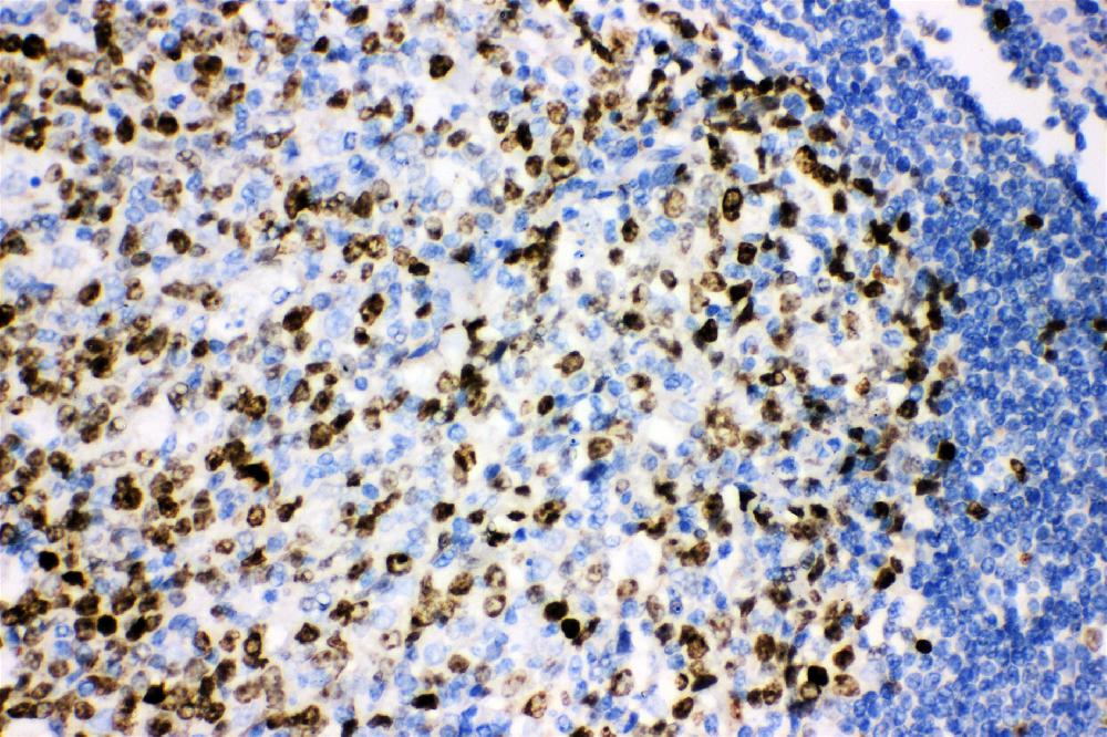
Figure 1. Immunohistochemical staining of paraffin-embedded Carcinoma of Human lung tissue using anti-MKI67 rat monoclonal antibody. (MA00254; heat-induced epitope retrieval by 10mM citric buffer, pH6.0, 120°C for 3min)
all(6)
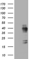
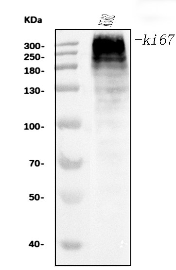
Figure 2. E.coli lysate (left lane) and E.coli lysate expressing human recombinant protein fragment corresponding to amino acids 1160-1493 of human MKI67 were separated by SDS-PAGE and immunoblotted with anti-MKI67.
all(6) 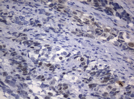
Figure 3. Immunohistochemical staining of paraffin-embedded Adenocarcinoma of Human endometrium tissue using anti-MKI67 rat monoclonal antibody. (MA00254; heat-induced epitope retrieval by 10mM citric buffer, pH6.0, 120°C for 3min)
all(6) 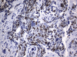
Figure 4. mmunohistochemical staining of paraffin-embedded Adenocarcinoma of Human breast tissue using anti-MKI67 rat monoclonal antibody. (MA00254; heat-induced epitope retrieval by 10mM citric buffer, pH6.0, 120°C for 3min)
all(6) 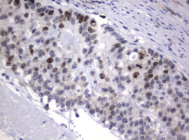
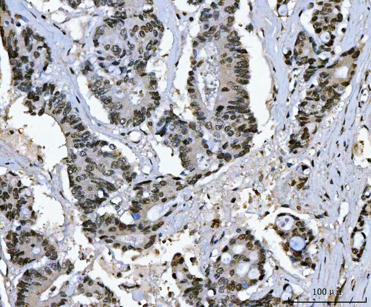
Figure 5. Immunohistochemical staining of paraffin-embedded Adenocarcinoma of Human colon tissue using anti-MKI67 rat monoclonal antibody. (MA00254; heat-induced epitope retrieval by 10mM citric buffer, pH6.0, 120°C for 3min)
all(6) 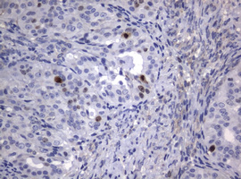
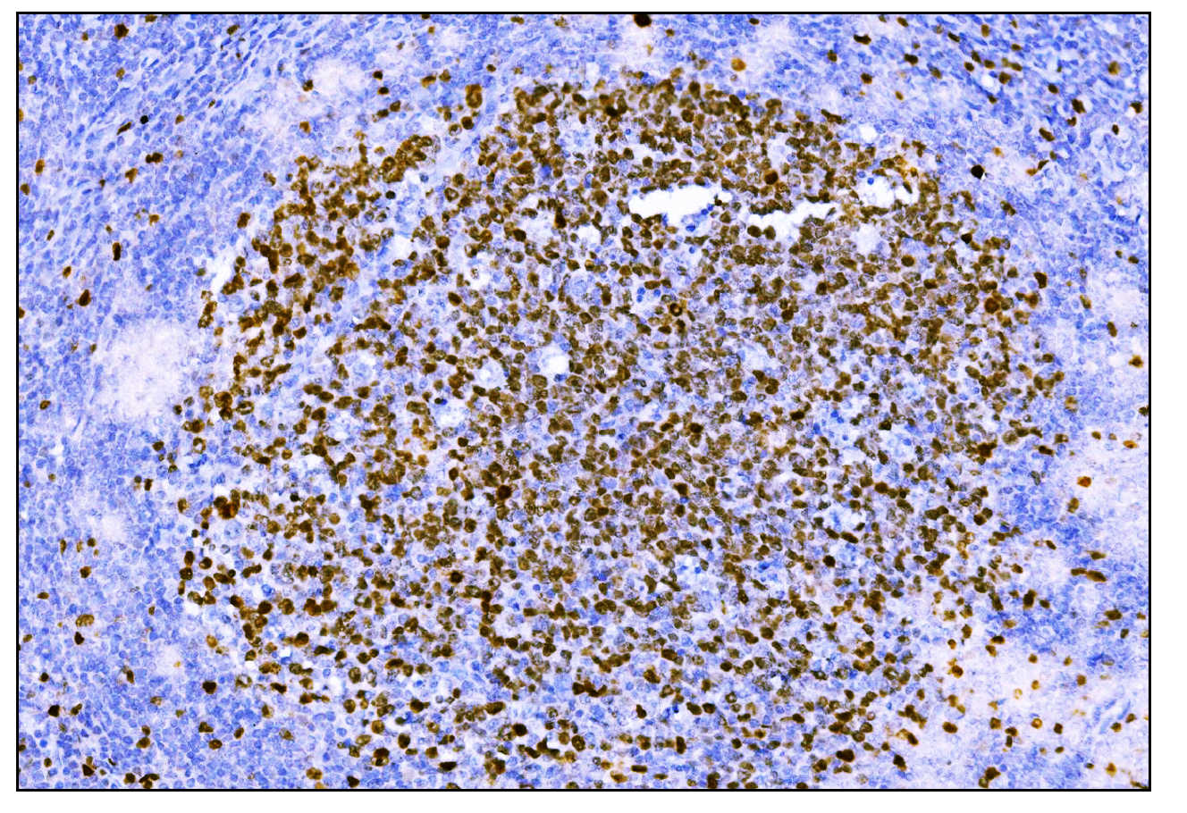
Figure 6. Immunohistochemical staining of paraffin-embedded Adenocarcinoma of Human ovary tissue using anti-MKI67 rat monoclonal antibody. (MA00254; heat-induced epitope retrieval by 10mM citric buffer, pH6.0, 120°C for 3min)
all(6) | Western blot (WB): | 1:1000 |
| Immunohistochemistry (IHC): | 1:150 |
Figure 1. Immunohistochemical staining of paraffin-embedded Carcinoma of Human lung tissue using anti-MKI67 rat monoclonal antibody. (MA00254; heat-induced epitope retrieval by 10mM citric buffer, pH6.0, 120°C for 3min)
Figure 2. E.coli lysate (left lane) and E.coli lysate expressing human recombinant protein fragment corresponding to amino acids 1160-1493 of human MKI67 were separated by SDS-PAGE and immunoblotted with anti-MKI67.
Figure 3. Immunohistochemical staining of paraffin-embedded Adenocarcinoma of Human endometrium tissue using anti-MKI67 rat monoclonal antibody. (MA00254; heat-induced epitope retrieval by 10mM citric buffer, pH6.0, 120°C for 3min)
Figure 4. mmunohistochemical staining of paraffin-embedded Adenocarcinoma of Human breast tissue using anti-MKI67 rat monoclonal antibody. (MA00254; heat-induced epitope retrieval by 10mM citric buffer, pH6.0, 120°C for 3min)
Figure 5. Immunohistochemical staining of paraffin-embedded Adenocarcinoma of Human colon tissue using anti-MKI67 rat monoclonal antibody. (MA00254; heat-induced epitope retrieval by 10mM citric buffer, pH6.0, 120°C for 3min)
Figure 6. Immunohistochemical staining of paraffin-embedded Adenocarcinoma of Human ovary tissue using anti-MKI67 rat monoclonal antibody. (MA00254; heat-induced epitope retrieval by 10mM citric buffer, pH6.0, 120°C for 3min)
 联系我们
联系我们027-67845390
 关注我们
关注我们
本司产品仅用于科研,不用于临床诊断和治疗
联系方式:027-67845390/1/2 技术支持:武汉丰网
© 1993-2025 Boster Biological Technology co.Itd E-mail:boster@boster.com
鄂ICP备05005548号-2
 鄂公网安备 42018502007312号

 积分商城
积分商城  购物车
购物车  登录/注册
登录/注册  您当前的位置:
  一键复制产品信息
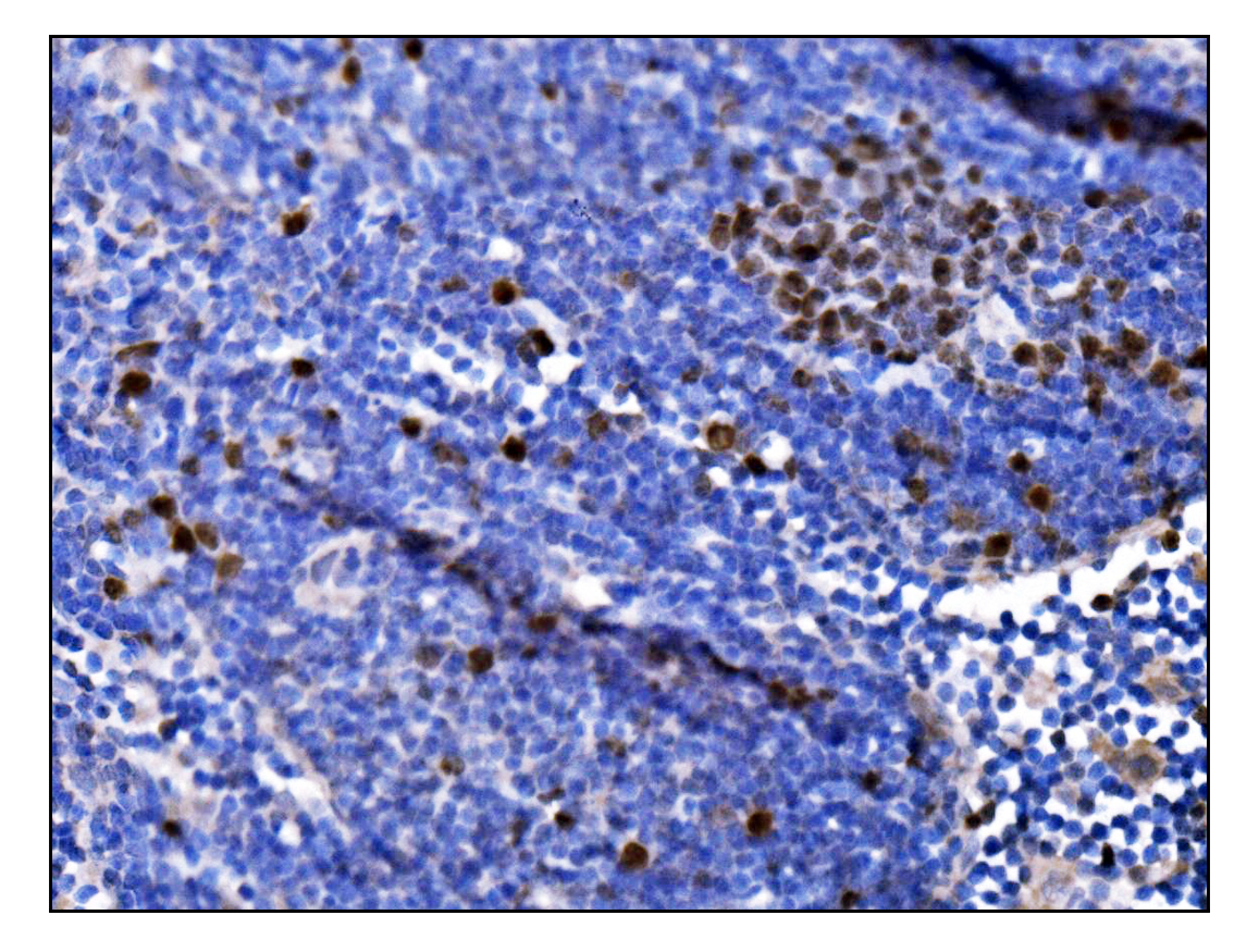
 成功添加到购物车
 微信客服
微信客服


 微信扫一扫立即咨询
