| Western blot (WB): | 1:500-2000 |
| Immunohistochemistry (IHC): | 1:50-400 |
| Immunocytochemistry/Immunofluorescence (ICC/IF): | 1:50-400 |
| Immunofluorescence (IF): | 1:50-400 |
| ELISA: | 1:100-1000 |
| (Boiling the paraffin sections in 10mM citrate buffer,pH6.0,or PH8.0 EDTA repair liquid for 20 mins is required for the staining of formalin/paraffin sections.) Optimal working dilutions must be determined by end user. | |
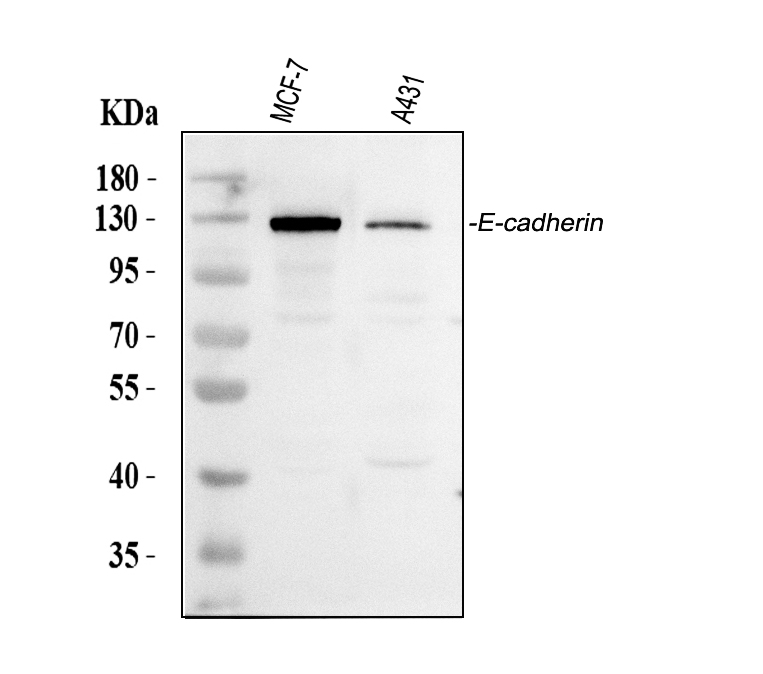
Western blot analysis of anti- CDH1/E-cadherin antibody (PB9561). The sample well of each lane was loaded with 30ug of sample under reducing conditions.
Lane 1: human MCF-7 whole cell lysates,
Lane 2: human A431 whole cell lysates.
Use rabbit anti- CDH1/E-cadherin 1:1000, probed with a goat anti-rabbit IgG-HRP secondary antibody. The signal is developed using an Enhanced Chemiluminescent detection (ECL) kit (Catalog#EK1002). A specific band was detected for CDH1/E-cadherin at approximately 130KD. The expected band size for CDH1/E-cadherin is at 97KD.

IHC analysis of E-cadherin/CDH1 using anti-E-cadherin/CDH1 antibody (PB9561).
E-cadherin/CDH1 was detected in a paraffin-embedded section of human placenta tissue. Biotinylated goat anti-rabbit IgG was used as secondary antibody. The tissue section was incubated with rabbit anti-E-cadherin/CDH1 Antibody (PB9561) at a dilution of 1:200 and developed using Strepavidin-Biotin-Complex (SABC) (Catalog # SA1022) with DAB (Catalog # AR1027) as the chromogen.
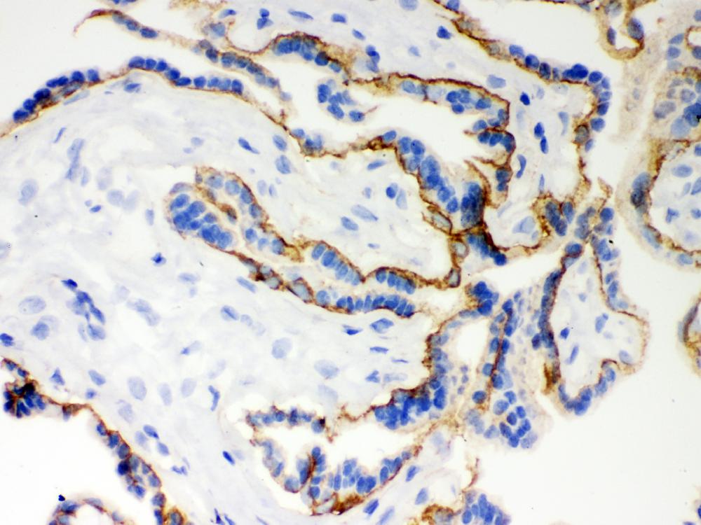
IHC analysis of E-cadherin/CDH1 using anti-E-cadherin/CDH1 antibody (PB9561).
E-cadherin/CDH1 was detected in frozen section of human placenta tissue. Biotinylated goat anti-rabbit IgG was used as secondary antibody. The tissue section was incubated with rabbit anti-E-cadherin/CDH1 Antibody (PB9561) at a dilution of 1:200 and developed using Strepavidin-Biotin-Complex (SABC) (Catalog # SA1022) with DAB (Catalog # AR1027) as the chromogen.
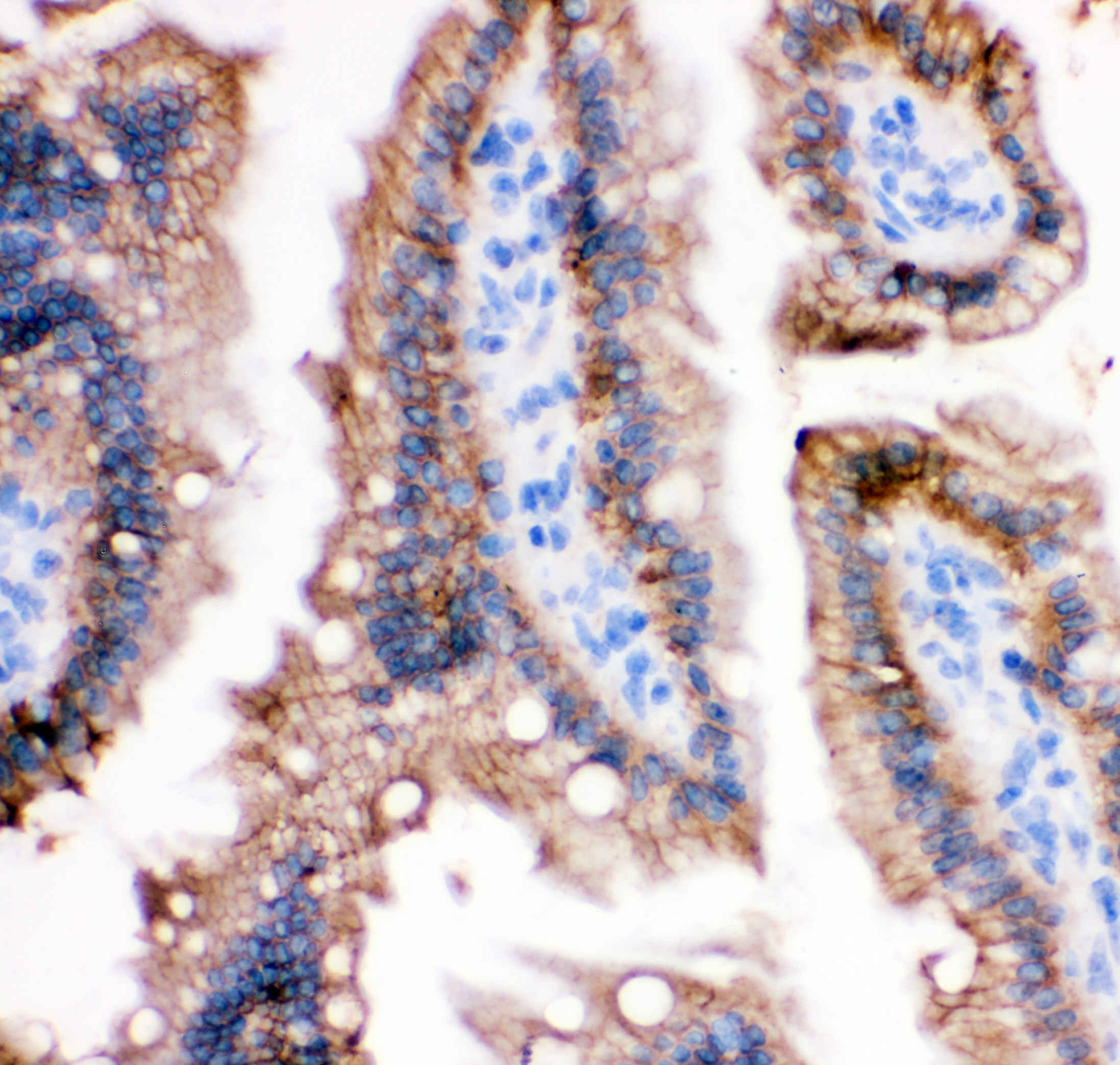
IHC analysis of E-cadherin/CDH1 using anti-E-cadherin/CDH1 antibody (PB9561).
E-cadherin/CDH1 was detected in a paraffin-embedded section of mouse intestine tissue. Biotinylated goat anti-rabbit IgG was used as secondary antibody. The tissue section was incubated with rabbit anti-E-cadherin/CDH1 Antibody (PB9561) at a dilution of 1:200 and developed using Strepavidin-Biotin-Complex (SABC) (Catalog # SA1022) with DAB (Catalog # AR1027) as the chromogen.
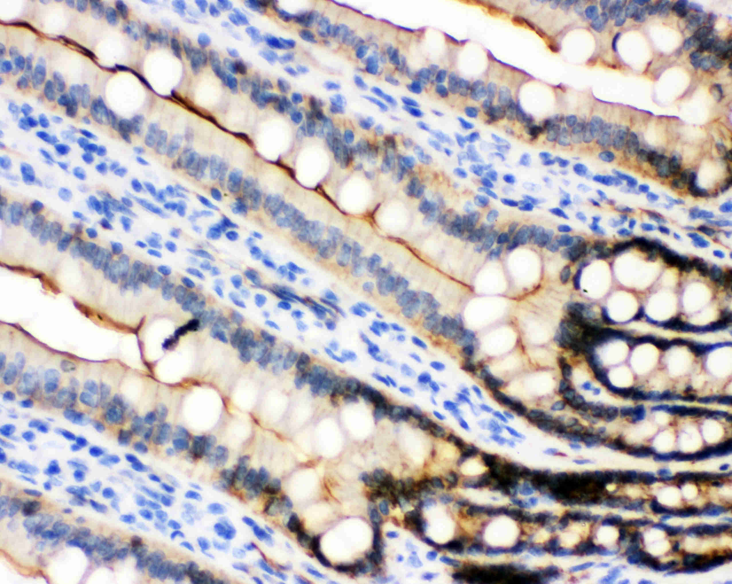
IHC analysis of E-cadherin/CDH1 using anti-E-cadherin/CDH1 antibody (PB9561).
E-cadherin/CDH1 was detected in a paraffin-embedded section of rat intestine tissue. Biotinylated goat anti-rabbit IgG was used as secondary antibody. The tissue section was incubated with rabbit anti-E-cadherin/CDH1 Antibody (PB9561) at a dilution of 1:200 and developed using Strepavidin-Biotin-Complex (SABC) (Catalog # SA1022) with DAB (Catalog # AR1027) as the chromogen.
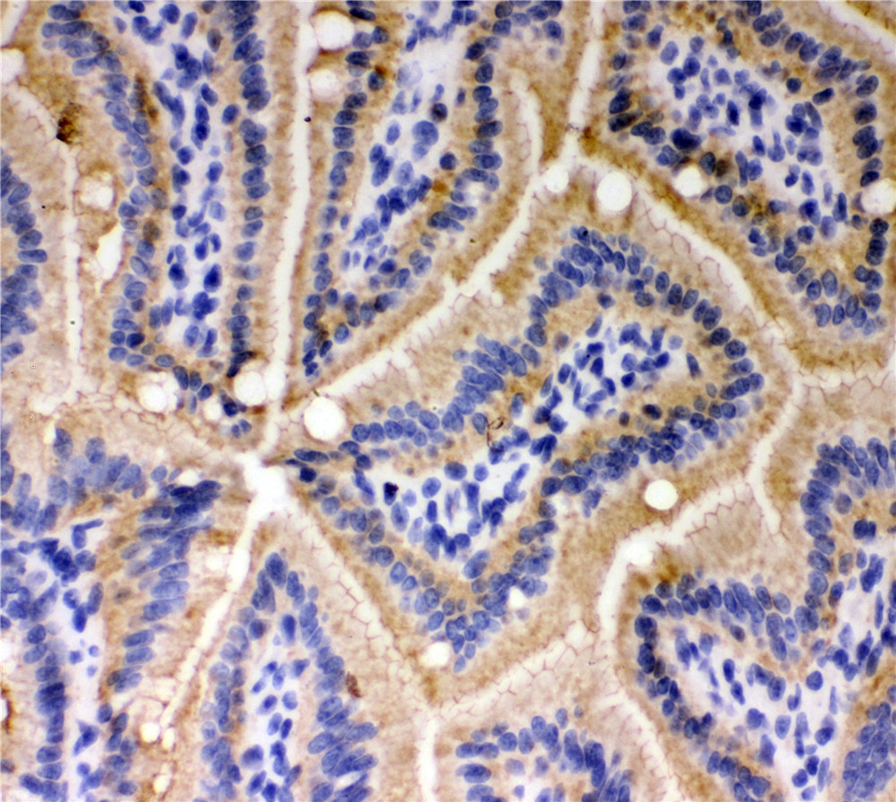
IHC analysis of E-cadherin/CDH1 using anti-E-cadherin/CDH1 antibody (PB9561).
E-cadherin/CDH1 was detected in frozen section of mouse intestine tissue. Biotinylated goat anti-rabbit IgG was used as secondary antibody. The tissue section was incubated with rabbit anti-E-cadherin/CDH1 Antibody (PB9561) at a dilution of 1:200 and developed using Strepavidin-Biotin-Complex (SABC) (Catalog # SA1022) with DAB (Catalog # AR1027) as the chromogen.

IHC analysis of E-cadherin/CDH1 using anti-E-cadherin/CDH1 antibody (PB9561).
E-cadherin/CDH1 was detected in frozen section of rat intestine tissue. Biotinylated goat anti-rabbit IgG was used as secondary antibody. The tissue section was incubated with rabbit anti-E-cadherin/CDH1 Antibody (PB9561) at a dilution of 1:200 and developed using Strepavidin-Biotin-Complex (SABC) (Catalog # SA1022) with DAB (Catalog # AR1027) as the chromogen.
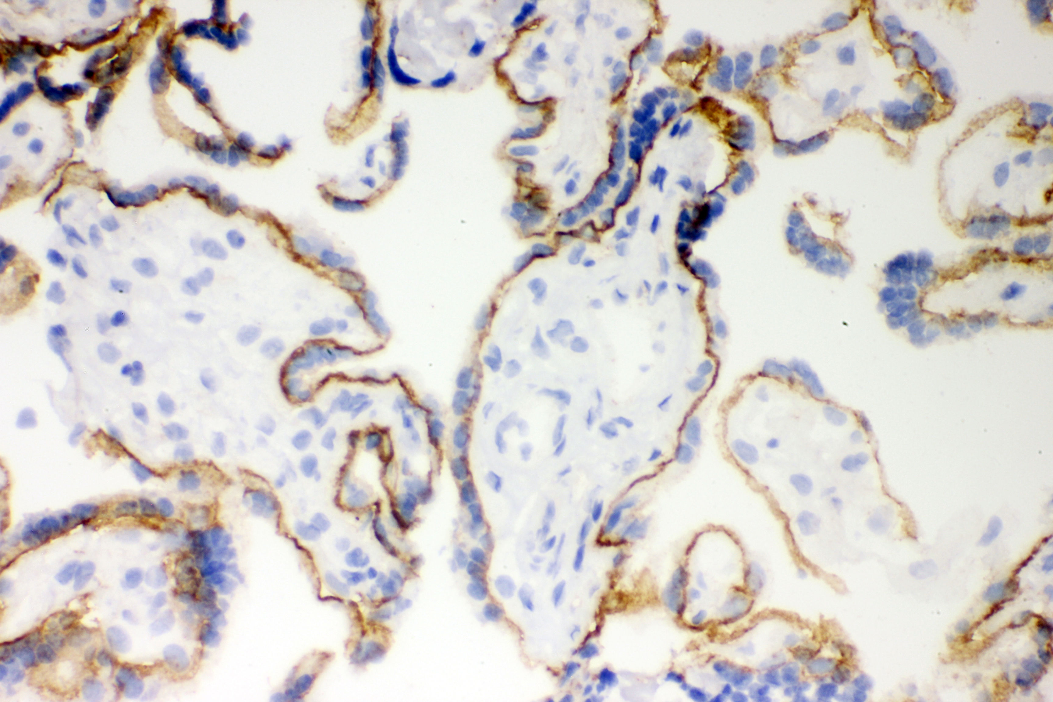
IHC analysis of E-cadherin/CDH1 using anti-E-cadherin/CDH1 antibody (PB9561).
E-cadherin/CDH1 was detected in frozen section of human placenta tissue. Biotinylated goat anti-rabbit IgG was used as secondary antibody. The tissue section was incubated with rabbit anti-E-cadherin/CDH1 Antibody (PB9561) at a dilution of 1:200 and developed using Strepavidin-Biotin-Complex (SABC) (Catalog # SA1022) with DAB (Catalog # AR1027) as the chromogen.
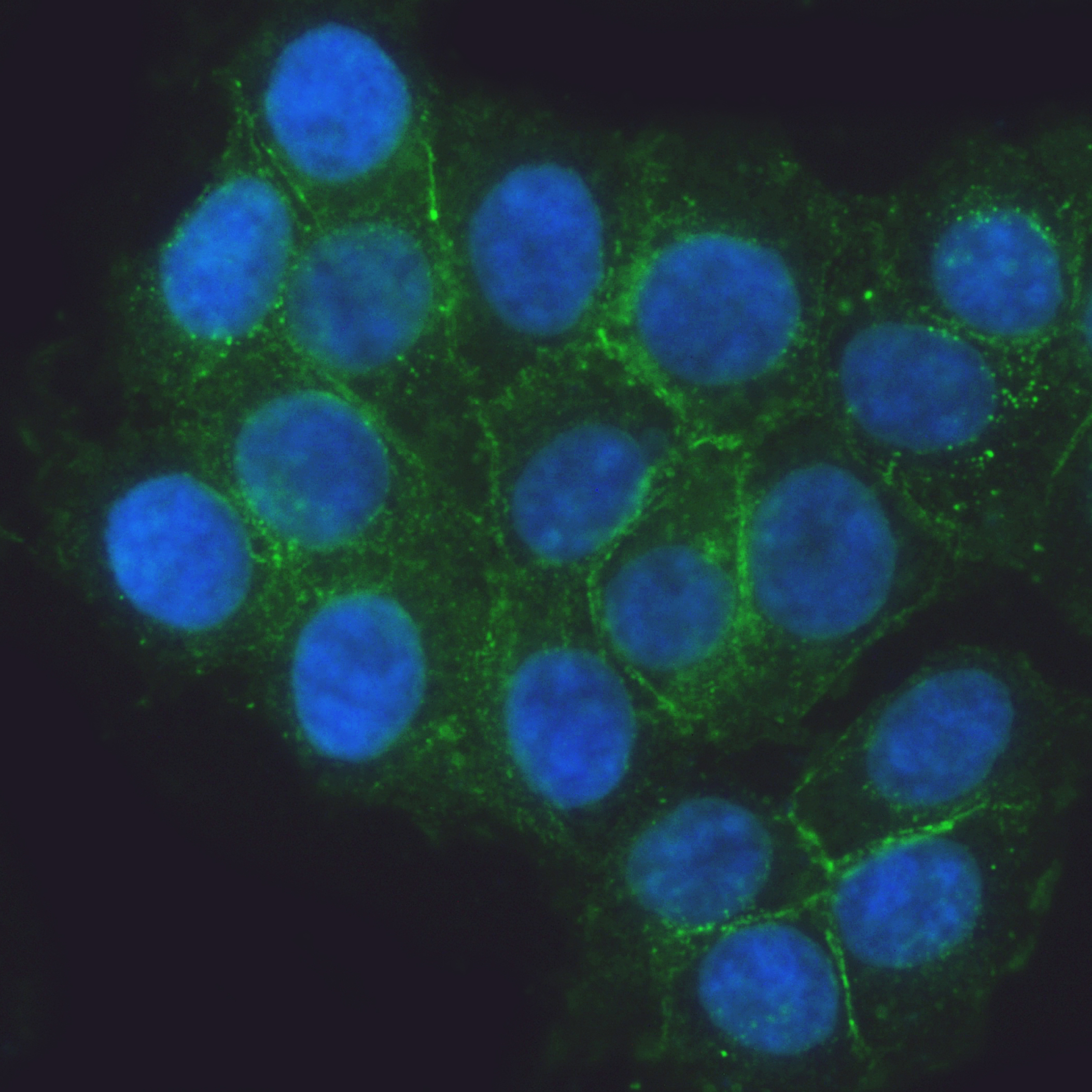
IF analysis of E-cadherin/CDH1 using anti-E-cadherin/CDH1 antibody (PB9561).
E-cadherin/CDH1 was detected in an immunocytochemical section of MCF-7 cells. The section was incubated with rabbit anti-E-cadherin/CDH1 Antibody (PB9561) at a dilution of 1:100. DyLight®488 Conjugated Goat Anti-Rabbit IgG (Green) (Catalog # BA1127) was used as secondary antibody. The section was counterstained with DAPI (Catalog # AR1176) (Blue).
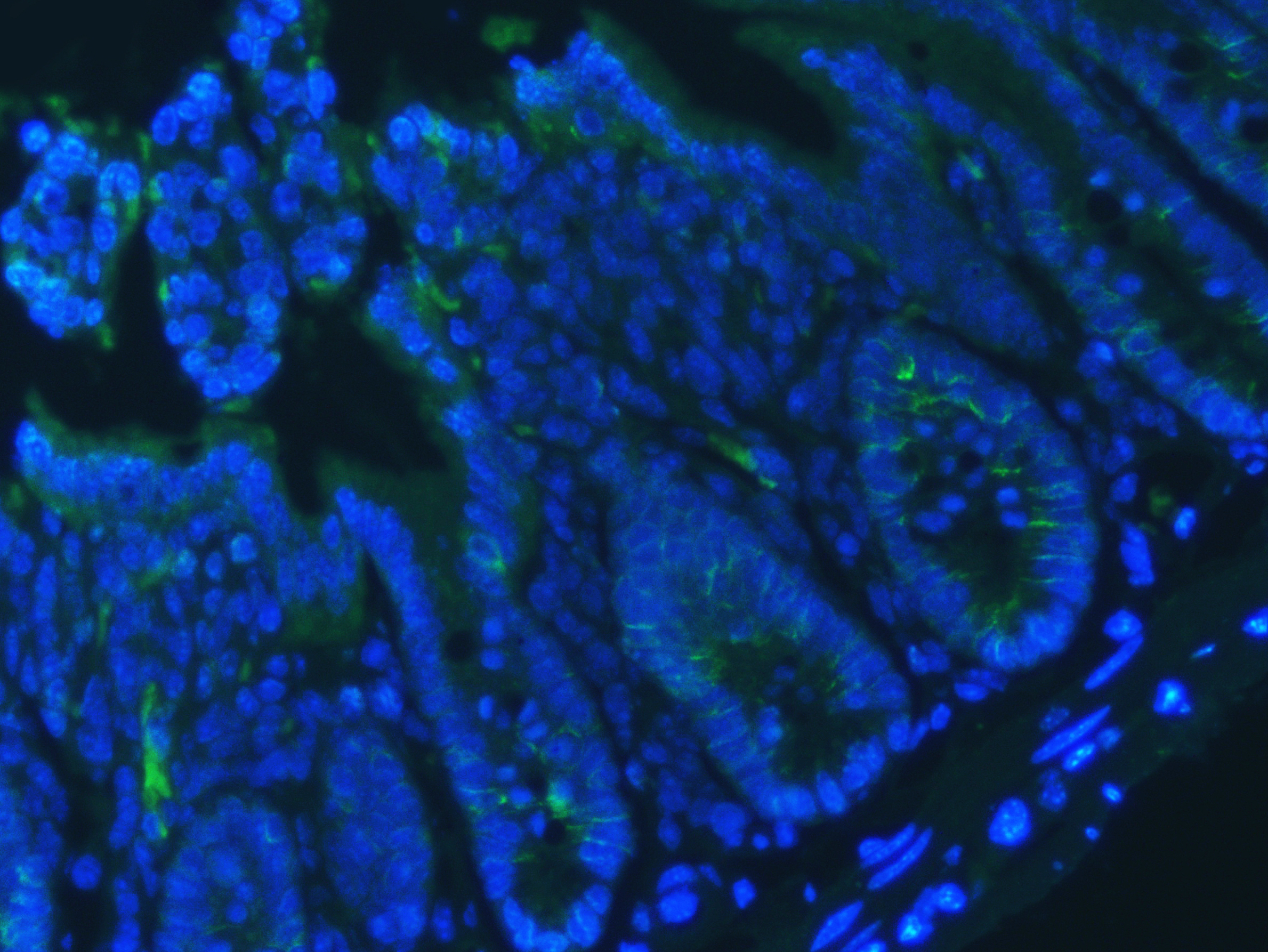
IF analysis using anti- E Cadherin antibody (PB9561). detected in paraffin-embedded section of mouse colon tissue. The tissue section were stained using the Dylight488-conjugated Anti-rabbit IgG Secondary Antibody (green) (Catalog # BA1127) and counterstained with DAPI (blue).
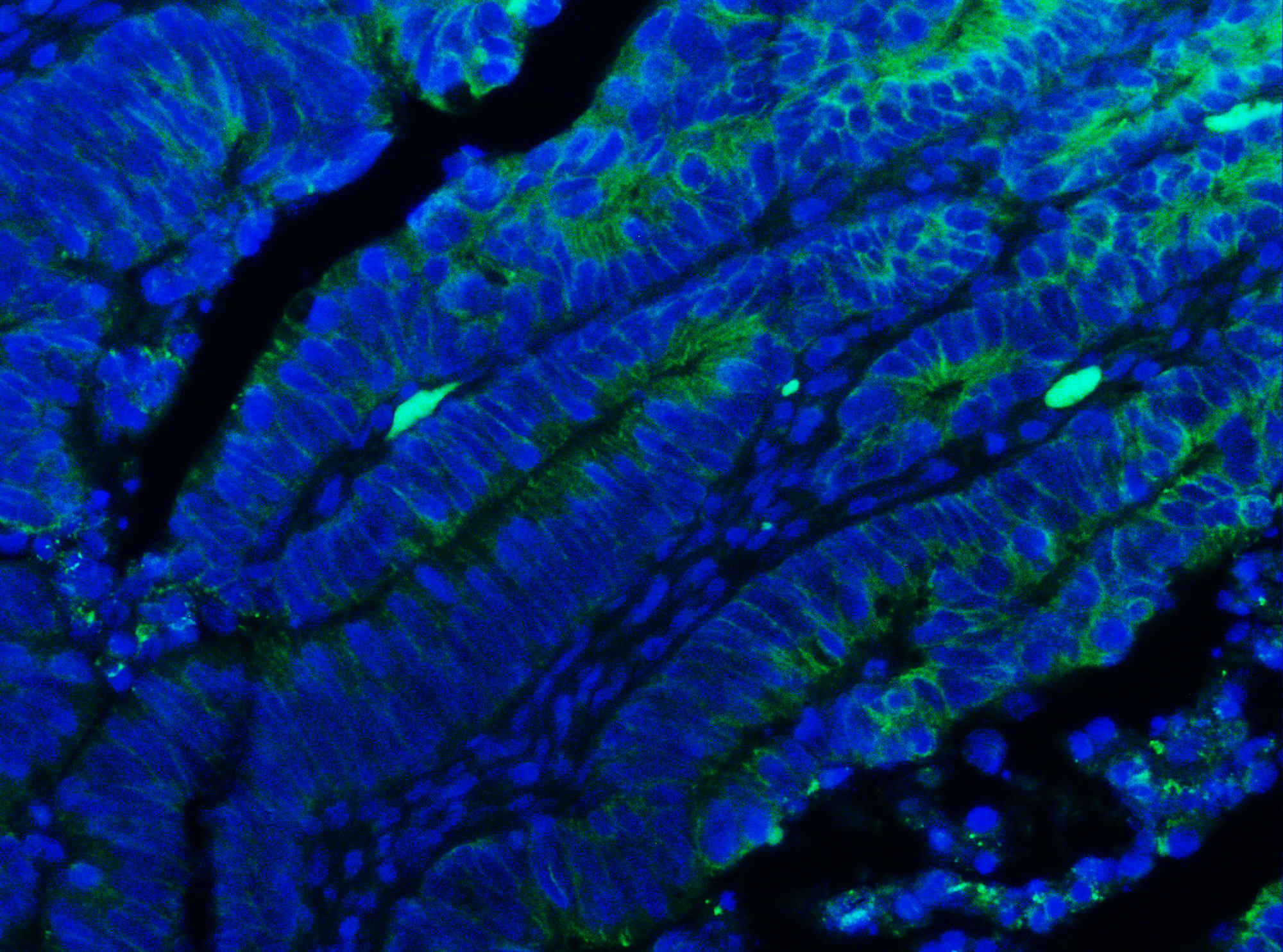
IF analysis using anti- E Cadherin antibody (PB9561). detected in paraffin-embedded section of human colon cancer tissue. The tissue section were stained using the Dylight488-conjugated Anti-rabbit IgG Secondary Antibody (green) (Catalog # BA1127) and counterstained with DAPI (blue).

Western blot analysis of anti- CDH1/E-cadherin antibody (PB9561). The sample well of each lane was loaded with 30ug of sample under reducing conditions.
Lane 1: human MCF-7 whole cell lysates,
Lane 2: human A431 whole cell lysates.
Use rabbit anti- CDH1/E-cadherin 1:1000, probed with a goat anti-rabbit IgG-HRP secondary antibody. The signal is developed using an Enhanced Chemiluminescent detection (ECL) kit (Catalog#EK1002). A specific band was detected for CDH1/E-cadherin at approximately 130KD. The expected band size for CDH1/E-cadherin is at 97KD.

IHC analysis of E-cadherin/CDH1 using anti-E-cadherin/CDH1 antibody (PB9561).
E-cadherin/CDH1 was detected in a paraffin-embedded section of human placenta tissue. Biotinylated goat anti-rabbit IgG was used as secondary antibody. The tissue section was incubated with rabbit anti-E-cadherin/CDH1 Antibody (PB9561) at a dilution of 1:200 and developed using Strepavidin-Biotin-Complex (SABC) (Catalog # SA1022) with DAB (Catalog # AR1027) as the chromogen.

IHC analysis of E-cadherin/CDH1 using anti-E-cadherin/CDH1 antibody (PB9561).
E-cadherin/CDH1 was detected in frozen section of human placenta tissue. Biotinylated goat anti-rabbit IgG was used as secondary antibody. The tissue section was incubated with rabbit anti-E-cadherin/CDH1 Antibody (PB9561) at a dilution of 1:200 and developed using Strepavidin-Biotin-Complex (SABC) (Catalog # SA1022) with DAB (Catalog # AR1027) as the chromogen.

IHC analysis of E-cadherin/CDH1 using anti-E-cadherin/CDH1 antibody (PB9561).
E-cadherin/CDH1 was detected in a paraffin-embedded section of mouse intestine tissue. Biotinylated goat anti-rabbit IgG was used as secondary antibody. The tissue section was incubated with rabbit anti-E-cadherin/CDH1 Antibody (PB9561) at a dilution of 1:200 and developed using Strepavidin-Biotin-Complex (SABC) (Catalog # SA1022) with DAB (Catalog # AR1027) as the chromogen.

IHC analysis of E-cadherin/CDH1 using anti-E-cadherin/CDH1 antibody (PB9561).
E-cadherin/CDH1 was detected in a paraffin-embedded section of rat intestine tissue. Biotinylated goat anti-rabbit IgG was used as secondary antibody. The tissue section was incubated with rabbit anti-E-cadherin/CDH1 Antibody (PB9561) at a dilution of 1:200 and developed using Strepavidin-Biotin-Complex (SABC) (Catalog # SA1022) with DAB (Catalog # AR1027) as the chromogen.

IHC analysis of E-cadherin/CDH1 using anti-E-cadherin/CDH1 antibody (PB9561).
E-cadherin/CDH1 was detected in frozen section of mouse intestine tissue. Biotinylated goat anti-rabbit IgG was used as secondary antibody. The tissue section was incubated with rabbit anti-E-cadherin/CDH1 Antibody (PB9561) at a dilution of 1:200 and developed using Strepavidin-Biotin-Complex (SABC) (Catalog # SA1022) with DAB (Catalog # AR1027) as the chromogen.

IHC analysis of E-cadherin/CDH1 using anti-E-cadherin/CDH1 antibody (PB9561).
E-cadherin/CDH1 was detected in frozen section of rat intestine tissue. Biotinylated goat anti-rabbit IgG was used as secondary antibody. The tissue section was incubated with rabbit anti-E-cadherin/CDH1 Antibody (PB9561) at a dilution of 1:200 and developed using Strepavidin-Biotin-Complex (SABC) (Catalog # SA1022) with DAB (Catalog # AR1027) as the chromogen.

IHC analysis of E-cadherin/CDH1 using anti-E-cadherin/CDH1 antibody (PB9561).
E-cadherin/CDH1 was detected in frozen section of human placenta tissue. Biotinylated goat anti-rabbit IgG was used as secondary antibody. The tissue section was incubated with rabbit anti-E-cadherin/CDH1 Antibody (PB9561) at a dilution of 1:200 and developed using Strepavidin-Biotin-Complex (SABC) (Catalog # SA1022) with DAB (Catalog # AR1027) as the chromogen.

IF analysis of E-cadherin/CDH1 using anti-E-cadherin/CDH1 antibody (PB9561).
E-cadherin/CDH1 was detected in an immunocytochemical section of MCF-7 cells. The section was incubated with rabbit anti-E-cadherin/CDH1 Antibody (PB9561) at a dilution of 1:100. DyLight®488 Conjugated Goat Anti-Rabbit IgG (Green) (Catalog # BA1127) was used as secondary antibody. The section was counterstained with DAPI (Catalog # AR1176) (Blue).

IF analysis using anti- E Cadherin antibody (PB9561). detected in paraffin-embedded section of mouse colon tissue. The tissue section were stained using the Dylight488-conjugated Anti-rabbit IgG Secondary Antibody (green) (Catalog # BA1127) and counterstained with DAPI (blue).

IF analysis using anti- E Cadherin antibody (PB9561). detected in paraffin-embedded section of human colon cancer tissue. The tissue section were stained using the Dylight488-conjugated Anti-rabbit IgG Secondary Antibody (green) (Catalog # BA1127) and counterstained with DAPI (blue).


